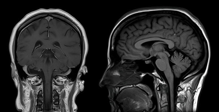
Magnetic resonance imaging (MRI) is a medical imaging technique that is used by doctors to visualize the internal structures of the body. MRI uses a powerful magnetic field, radio waves, and computers to create detailed pictures of the inside of the body.
MRI is regarded as one of the safest, low-risk medical imaging techniques a patient could have. Unlike X-rays and CT scans, MRI scans require no ionizing radiation. Instead, it uses strong magnetic fields and radio waves to create clear images of organs and tissues.
MRI can be used to image almost any part of the body, and it is commonly used in neurology, as it can visualize the brain and spinal cord. Results of the MRI can reveal to neurologists aneurysms, abscesses, plaque, tumors, and injuries.
In this article, we will talk about how MRI imaging works.
MRI Procedure
The MRI procedure begins by having you lie on a table that slides into a large tunnel-like machine called an MRI scanner. You will be asked to stay still during the scan, so that clear images can be obtained. The scanner emits loud thumping noises during scanning, which may last up to 30 minutes, depending on what area is being imaged. Earplugs are provided to help reduce noise levels for the patient, if needed.
Once you are positioned inside the scanner, coils located around you will emit radio waves directed at the specific area of interest, while simultaneously measuring very faint signals coming from your body in response. A computer then processes this information and produces detailed cross-sectional images (“slices”) through whatever part of your body was scanned. These slices can then be viewed as individual pictures or stacked together like pieces of a puzzle to give a 3D image.
In some cases, you may need an IV line inserted into your vein before beginning an MRI exam, so that contrast material can be injected, if needed. This contrast material contains gadolinium, which helps make certain structures more visible and clear in images.
Afterward, you can return home and resume normal activities immediately. Your doctor will interpret the results of your scan and discuss them with you at a follow-up appointment.
MRI Imaging in Howell, MI
Associates in Neurology is a top neurology practice in Southeast Michigan, with locations in Howell, Farmington Hills, and Novi. Our board-certified neurologists use magnetic resonance imaging to evaluate our patients’ symptoms and make diagnoses. We use MRI to diagnose stroke, multiple sclerosis, aneurysms, traumatic brain injuries, spinal cord disorders, and other neurological conditions.
If you need a brain or spinal cord MRI, choose us as your provider. Our doctors are recognized in the field of neurology and provide only the best and most compassionate care to patients. For any questions or to schedule a consultation, call our office today at (248) 478-5512. You can also send an appointment request by using our convenient online form. We look forward to serving you!


